04:37 19 January
in by
adminadam
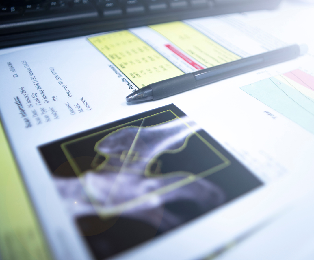
[vc_row type="grid" border_color="#eaeaea" padding_top="50" padding_bottom="50"][vc_column width="2/3"][vc_tta_tour shape="round" color="sky" active_section="1"][vc_tta_section title="What is BMD" tab_id="1515724954892-df2435f6-1e9d"][vc_column_text]Using Dual Energy X-ray Absorption (DEXA), this examination is used to measure Bone Mineral Density (BMD). The most common cause for loss of bone density is osteoporosis.
Given that women lose bone from about the age of 45 (linked with Oestrogen production falling), a Bone Density Scan that enables a like for like comparison over time is useful to alert patients and provide time for them to minimise risks of Osteoporosis.
Information about Osteoporosis can be found at the Osteoporosis Australia website here.
The Health Direct Website provided by the Australian...
04:34 10 November
in by
ncradgroup
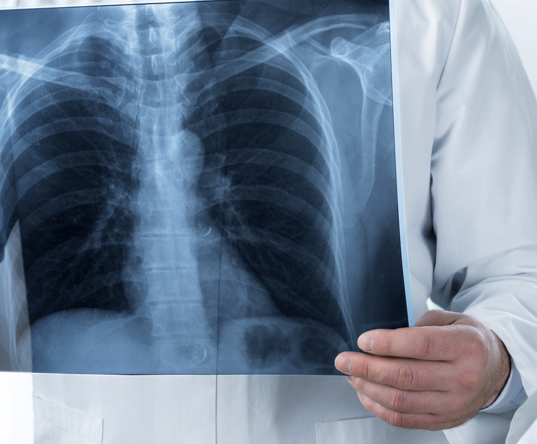
[vc_row type="grid" border_color="#eaeaea" padding_top="50" padding_bottom="50"][vc_column width="2/3"][vc_video link="https://youtu.be/aIx8uZl1-6U"][vc_empty_space][vc_tta_tour shape="round" color="sky" active_section="1"][vc_tta_section title="What are Xrays" tab_id="1515633672130-3d8cbeca-70a5"][vc_column_text]The test relies on the fact that different parts of the body attenuate (stop) X-rays better than others. X-rays are ionising radiation, generated by an X-ray tube.
The rays are controlled by shielding down a narrow beam, directed towards the part of the body being examined. On the opposite side of the body, an x-ray film is positioned in the path of the x-rays, and is "exposed".
The X-ray film is then processed and an image generated. Where the X-ray passes through the body easily, that part of the...
04:33 10 November
in by
ncradgroup
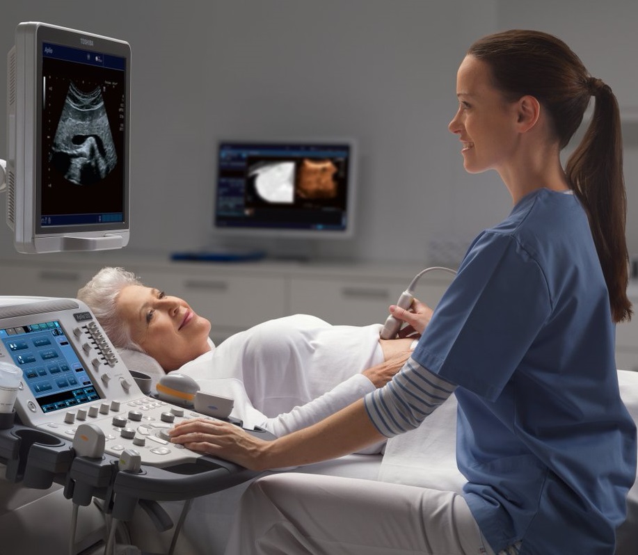
[vc_row type="grid" border_color="#eaeaea" padding_top="50" padding_bottom="50"][vc_column width="2/3"][vc_video link="https://youtu.be/wxJdMe6U9YU"][vc_empty_space][vc_tta_tour shape="round" color="sky" active_section="1"][vc_tta_section title="What is Ultrasound" tab_id="1515627504923-3df9f52f-5ddd"][vc_column_text]An ultrasound scan is a procedure that uses high frequency sound waves to generate images of internal body structures. Ultrasound does not use ionising radiation so is considered very safe.
To acquire the images water-based gel is applied to your skin and then a hand-held ultrasound transducer is moved over the area of interest.
The ultrasound examination is performed by our highly trained Sonographers who then forward the images to our specialist doctors. The Radiologist then prepares a report for your referring practitioner.[/vc_column_text][/vc_tta_section][vc_tta_section title="Preparation" tab_id="1515627639848-2e7cdc77-ce81"][vc_column_text]
Diagnostic Ultrasound examinations often have...
04:33 10 November
in by
ncradgroup
[vc_row type="grid" border_color="#eaeaea" padding_top="50" padding_bottom="50"][vc_column width="2/3"][vc_tta_tour shape="round" color="sky" active_section="1"][vc_tta_section title="About" tab_id="1515731540757-87f72e95-98a0"][vc_column_text]
Mammography
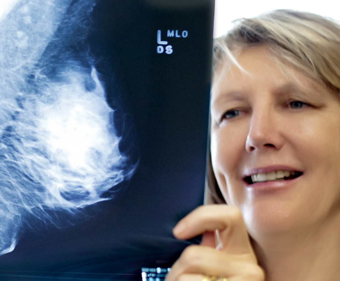
Mammograms involve a low dose X-ray examination of the Breasts. Mammography plays an important part in the early detection of Breast Cancer, before there are any signs or symptoms of the disease. A Mammogram can be either 2D or 3D.
A 3D Mammogram captures a series of thin ‘layers’ (around 1mm thick) through the breast providing greater detail.
3D Mammography can demonstrate early invasive breast cancers more clearly than 2D Mammography alone. 3D Mammograms may be more valuable for those with dense breast tissue or implants.
2D Mammogram capture images in 2 dimensions....
04:32 10 November
in by
ncradgroup
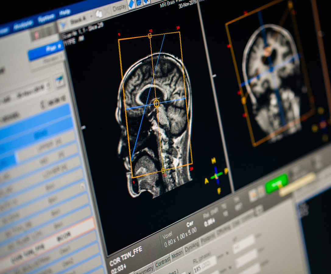
[vc_row type="grid" border_color="#eaeaea" padding_top="50" padding_bottom="50"][vc_column width="2/3"][vc_video link="https://youtu.be/Cmt8xqfCzbI"][vc_empty_space][vc_tta_tour shape="round" color="sky" active_section="1"][vc_tta_section title="What is MRI" tab_id="1515728313177-0218da86-5a37"][vc_column_text]Magnetic Resonance Imaging (MRI) is a method of looking inside the body without using surgery or x-rays. The MR scanner is a large doughnut shaped magnet open at both ends. It uses a strong magnetic field, radio waves and a computer to produce clear pictures of the human body. This technology is important because MRI scans can demonstrate to your doctor the difference between healthy and diseased tissue.[/vc_column_text][/vc_tta_section][vc_tta_section title="Preparation" tab_id="1515728313213-bb6cf8a9-5d69"][vc_column_text]There are certain patients on whom we cannot perform the test. That is why we ask all patients...
04:32 10 November
in by
ncradgroup
[vc_row type="grid" border_color="#eaeaea" padding_top="50" padding_bottom="50"][vc_column width="2/3"][vc_tta_tour shape="round" color="sky" active_section="1"][vc_tta_section title="About" tab_id="1515726901871-88182317-f700"][vc_column_text]Our radiologists experienced in undertaking various proceedures and interventional Radiology which include:
Injections
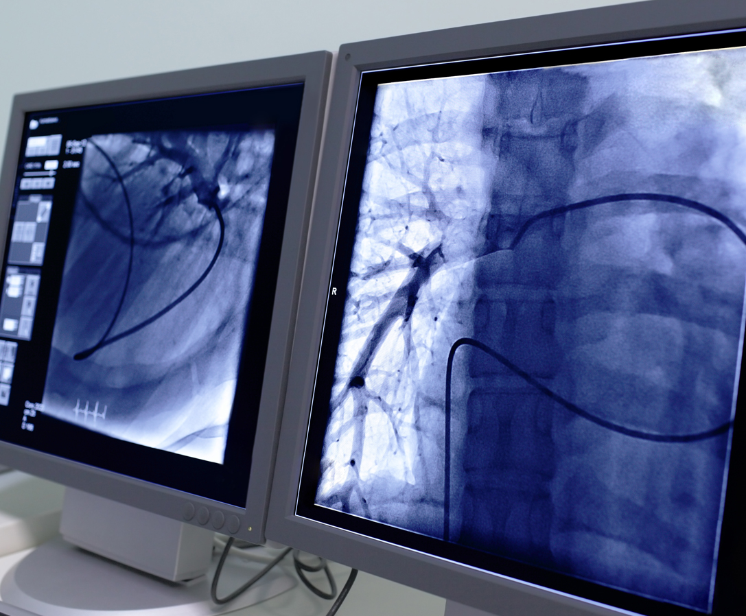
Injections are common for areas such as nerve roots and shoulders. A corticosteroid injection around nerve roots, for example, may alleviate the pain by reducing inflammation of the nerve. If the pain is suspected to come from a particular root, but it is not certain which one (especially in older people who may have root compression as a result of arthritis at a number of spinal levels), blocking the root with anaesthetic confirms or rules out a particular root...
04:31 10 November
in by
ncradgroup
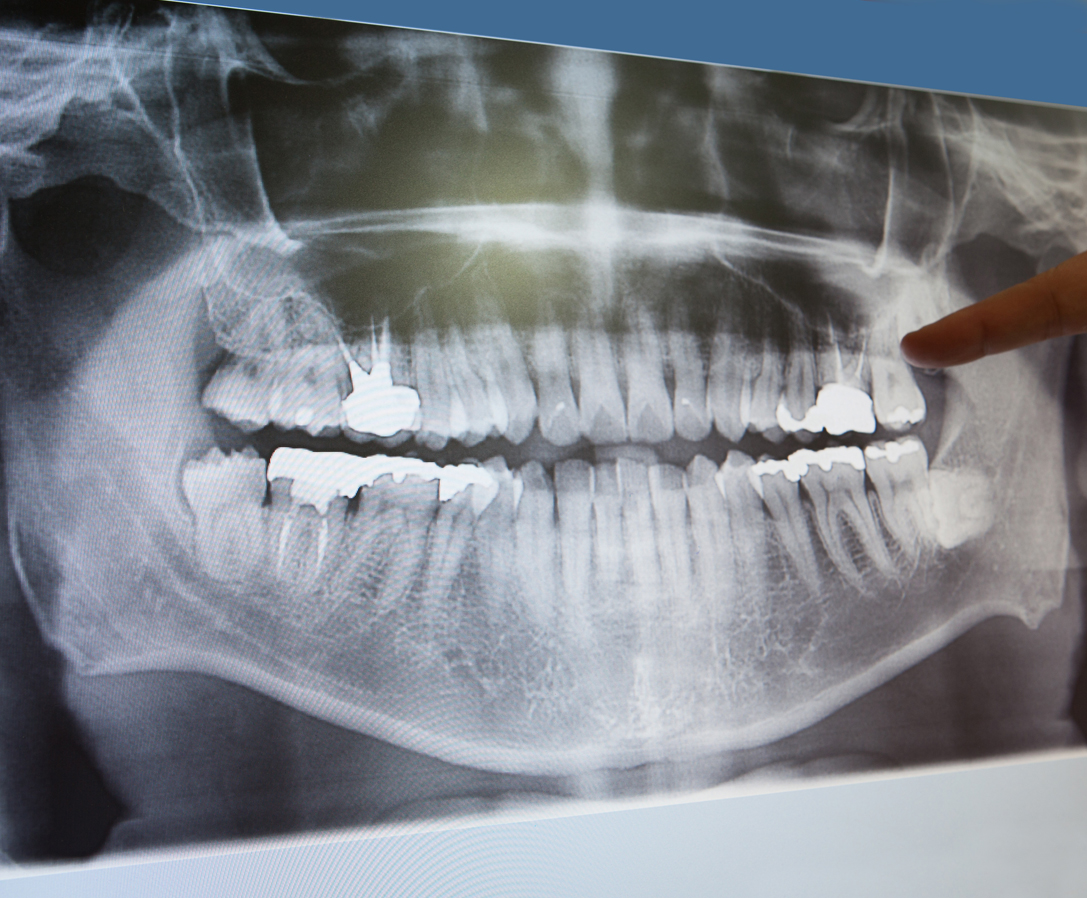
[vc_row type="grid" border_color="#eaeaea" padding_top="50" padding_bottom="50"][vc_column width="2/3"][vc_tta_tour shape="round" color="sky" active_section="1"][vc_tta_section title="What is Dental Imaging" tab_id="1515726143939-713c66f3-b514"][vc_column_text]Dental Imaging involves not only looking at the teeth but also the upper and lower jaws, and often the entire face. Your dentist/oral surgeon may also require information on bone growth when planning surgical procedures for younger people.
The basic tools of dental imaging are the OPG (Orthopantomogram) and Cephalometry. These x-rays require specialised x-ray machines. An OPG (“orthopantomogram”) gives a panoramic view of the mouth, giving information on the teeth and the bones of the upper and lower jaw. Cephalometry is used to obtain measurements and determine...
04:31 10 November
in by
ncradgroup
[vc_row type="grid" border_color="#eaeaea" padding_top="50" padding_bottom="50"][vc_column width="2/3"][vc_video link="https://youtu.be/zjRgpGL4zj4"][vc_empty_space][vc_tta_tour shape="round" color="sky" active_section="1"][vc_tta_section title="What are CT scans" tab_id="1515564396093-94961b73-1382"][vc_column_text]Computed Tomography (CT) Scan is used to look at the internal structures of the body. Both soft tissues (eg brain, liver, kidneys, lungs) and bone can be seen. The images are cross-sections of the body, but the CT computer can generate a great variety of images, depending on what the doctor is looking for.
For more detailed information on CT examinations click on the tabs or research the Inside Radiology Industry website here.[/vc_column_text][/vc_tta_section][vc_tta_section title="Types of scans" tab_id="1515565347938-81bb742f-33a7"][vc_column_text]CT Scans are commonly requested for the following areas of the...


