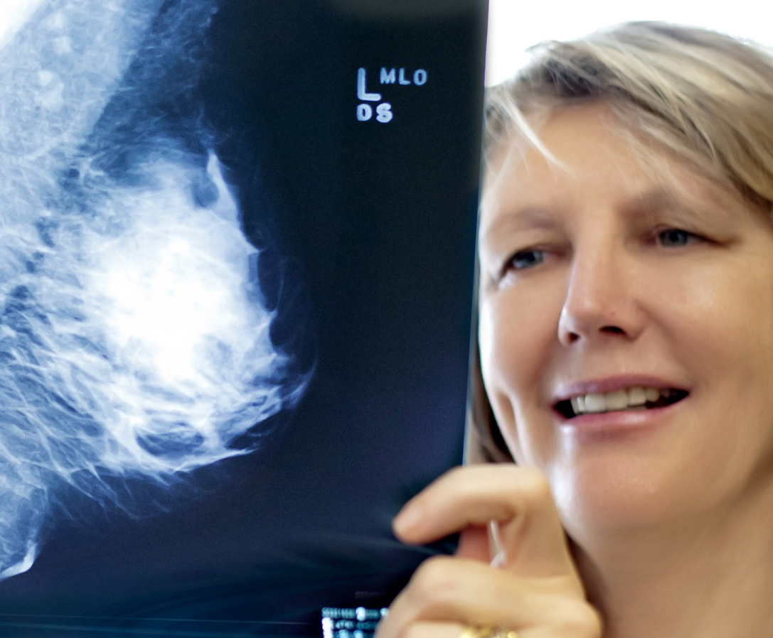
Mammography

Breast Imaging - Mammography - Tomography

Mammography

Mammograms involve a low dose X-ray examination of the Breasts. Mammography plays an important part in the early detection of Breast Cancer, before there are any signs or symptoms of the disease. A Mammogram can be either 2D or 3D.


A 3D Mammogram captures a series of thin ‘layers’ (around 1mm thick) through the breast providing greater detail.


3D Mammography can demonstrate early invasive breast cancers more clearly than 2D Mammography alone. 3D Mammograms may be more valuable for those with dense breast tissue or implants.


2D Mammogram capture images in 2 dimensions. This may be appropriate for those patients with non-dense breast tissue and/or where a lump may be larger.

Breast Ultrasound

Not all breast cancers can be detected by mammography alone and referring Doctors usually request a Mammogram in conjunction with an Ultrasound. Ultrasound uses sound waves and can find different types of lesions to Mammograms.


US of the breast is a real time examination; a patient can indicate the location of a lump and it can be immediately viewed and correlated by the Radiologist with Mammography.


Ultrasound is also used to assess lumps seen on Mammography and to determine if they are solid or cystic.


Microcalcification, which can be a sign of early cancer, is generally not visible on Ultrasound and is usually found with Mammography.


3D Mammogram technology is available at Lismore Women’s Imaging, Ballina  and Grafton.


Breast Biopsy

Biopsies are a specialist radiological procedure performed by the Radiologist using Ultrasound to obtain a sample of a lesion for a Pathologist to examine.


A biopsy of a breast lesion under 6mm in size may result in removing most of the lesion and therefore the site of the lesion could be difficult to locate in the future. A Radiologist may recommend the placement of a breast tissue marker (breast clip) to ensure the site of the lesion could be located again if needed.

Implants

If you have an implant, you may be concerned about the impact of a Mammogram in terms of potential leakage/rupture. Having an implant makes a Mammogram no less important. Mammograms are generally safe if you have an implant unless there is already an issue with the implant.


The amount of compression used in Mammograms is less when imaging patients with implants, to reduce any risk of rupture. The implants can obscure some of the breast tissue on Mammography but this is minimised with 3D Mammograms as they reduce hidden tissue to a minimum.


Do Men have Mammograms?


Yes, Men can and often do have Mammograms. Men may have “lumps” in the breast, as women do. Most often, as with women, these lumps are not cancer, but caused by hormonal changes in the body. Rarely, a male breast lump may be malignant. One percent of all breast cancers occur in men.


For information for women with implants click here


For more information from Inside Radiology click here

No perfume, deodorant, lotions (eg moisture cream) or talcum powder as this can show up on the x-ray and impact the accuracy of the image.


Finally, wear a two piece outfit that allows you to continue to wear the bottom section (eg trousers or skirt) but for ease of examination allows the top section (shirt) to be removed. This will assist with providing privacy and warmth.


If you experience significant breast discomfort during your menstrual period, it may be best not to schedule your mammogram during this time. One week after you period is best, unless the examination is urgent.


Eat and drink normally, and continue to take you usual medications.


It is very important to bring previous films to your appointment – even if your previous Mammogram was done by us. This is to support a thorough analysis of your examination.

The test may be uncomfortable for some people, but should not be the cause of terrible pain. You should understand that a compression plate is placed on the breast for very good reasons: to allow x-rays to pass through a thinner region which will decrease the x-ray dose and increase the accuracy of the test, and to move different parts of the breast off other parts of the breast, thus revealing hidden structures.


In 2D Mammography, two standard views are obtained: one from the top (“cranio-caudal” or CC) and one from the side (“medio-lateral oblique” or MLO).


Depending on what is found from these two, other specialised views may be required. For example, if the radiologist wants to better look at a small area of calcification, a magnified view can be obtained. It is important that you should not be shocked or worried that other mammograms are obtained. Most of the time, the radiologist is trying to either get a better look at something, or trying to move overlapping parts of the breast away from each other. Having extra views taken is NOT necessarily a bad sign.


In 3D Mammography the x-ray arm sweeps in a slight arc over the breast taking a series of images at various angles in just seconds.


3D Mammography can also be used for highly accurate placement of localisation wire in less time making for less discomfort for the patient and a high level of accuracy.


A Mammogram examination takes about 20 minutes. The actual image capture time for 3D is just a few seconds more compared with 2D Mammography.

3D Mammography Referrer Information


Requesting a 3D Mammogram vs 2D

3D Mammograms can be requested in the same way as 2D Mammograms and need to meet the same medicare requirements.  Since not all NCRG locations have the 3D Mammogram technolocy in place, the Mammogram done will be per the capabilities of the location. Currently, 3D Mammogram is available in Lismore,  Ballina, Chatswood and Grafton.


Please indicate 3D Views’ or ‘Tomosynthesis/Tomography’ on the referral if you specifically want your patient to obtain a 3D study.

The Government uses rules to determine if rebates offered by Medicare and the Department of Veterans Affairs can be accessed by patients. For patients to be eligible for a Medicare rebate the following information must be present and stated on the referral.


Requirements

Requesting doctors must include relevant clinical indications/history for all mammography (2D and 3D)  procedures otherwise patients are not eligible for a Medicare rebate.
For patients to be eligible for a Medicare rebate you must include your reasons to suspect malignancy in the breasts because of:


  • The past occurrence of breast malignancy in the patient or in a member of the patient’s family when a blood relative; or
  • Symptoms or indications of malignancy found on an examination of the patient by a medical practitioner.

A mammogram which has ‘routine’ or ‘screening’ written on it, will not quality for a Medicare rebate.


For 3D Mammograms examinations, the same Medicare eligibility rules apply. When booking a Mammogram appointment you must indicate which Mammogram (2D or 3D) is being requested as not all branches have the same Mammogram technology in place and it will affect which branch the appointment is booked at. It also affects the medicare rules so 3D must be written onto the referral when required.

Service Availability

Mammography
Ballina, Grafton, Mammo, Services, Women’s Imaging
Ballina
BMD, Branches, CT, Interventions, Mammo, MRI, NCR, OPG, X-Ray
Grafton
BMD, Branches, CT, CVI, DXA Body Composition, Interventions, Locations, Mammo, MRI, OPG, Screening, Ultrasound, X-Ray